


Anna C. Frick, MD
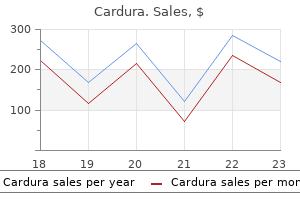
Cardura dosages: 4 mg, 2 mg, 1 mg
Cardura packs: 30 pills, 60 pills, 90 pills, 120 pills, 180 pills, 270 pills, 360 pills

The medial walls of the maxillary sinuses and the inferior ethmoid air cells are eroded arrhythmia svt cardura 4 mg buy with mastercard. There is near-complete opacification of ethmoid air cells, extensive erosion of ethmoid septa, and new bone formation (arrowheads; a). The maxillary sinuses are completely opacified, the medial walls are eroded, and the remaining walls exhibit hyperostosis (arrows; b). Similar changes of opacification and wall thickening and sclerosis are also seen in the sphenoid sinuses. The left cavernous sinus has an abnormally thick enhancing component extending to the foramen ovale (arrowheads; a). The thickening extends posteriorly along the left tentorial leaflet (black arrows; b) as well as anteriorly through the superior orbital fissure (white arrows; b). Pathology affecting these regions was combined in one chapter, rather than discussed in isolation, because of their close anatomic relationship and the consequent overlap in disease or complications affecting adjacent anatomic regions. Imaging has an essential and central role in the evaluation of pathology affecting these regions as well as preoperative staging and treatment planning in cases of neoplastic disease. In certain cases, imaging enables accurate diagnosis and identification of benign disease or "no touch" lesions that do not require any intervention. When evaluating malignant disease, certain imaging features can narrow the differential or occasionally suggest a specific histologic type. However, in general, the final diagnosis of malignant disease is based on biopsy and histopathology. For this group of diseases, one of the most important contributions of imaging is to accurately evaluate the extent of disease and upstage the initial clinical assessment by identifying disease spread to regions that cannot be reliably evaluated on clinical examination. Imaging identification of important anatomic variants is also important for preoperative planning and avoiding potential complications, particularly when minimally invasive endoscopic approaches are used. In this regard, the aim of this chapter was not only to review the imaging appearances of common and uncommon pathologies but also discuss relevant anatomic variants or spread patterns that are important and help optimize patient management. The specific pathways involved with perineural spread depend on the anatomic site and extent of the primary lesion: carefully evaluate the major neural pathways supplying the area of involvement by the primary tumor. Malignant tumors and aggressive infections may not always be distinguishable by imaging features alone; clinical information including patient risk factors and presentation can however be helpful in narrowing the differential diagnosis; if in doubt, biopsy may be required. An assessment of the relationship between the maxillary sinus floor and the maxillary posterior teeth root tips using dental cone-beam computerized tomography. It is important to correctly recognize developmental variants and benign lesions in the skull base to avoid unnecessary invasive procedures or biopsy. Computed tomographic imaging to determine the frequency of anatomical variations in pneumatization of the ethmoid bone. Endoscopic surgery in the treatment of crista galli pneumatization evolving with localizated frontal headaches. Lateral lamella of the cribriform plate: software-enabled computed tomographic analysis and its clinical relevance in skull base surgery.
It rotates the tibia medially on the femur or pulse pressure emedicine 1 mg cardura buy, when the tibia is fixed, rotates the femur laterally on the tibia. It retracts the posterior horn during lateral rotation and continuing flexion, via its attachment to the lateral meniscus, and so prevents traumatic compression. It provides dynamic stability to the posterolateral part of the knee by preventing excessive lateral rotation of the tibia, partly by its direct action, but more significantly by tensing the popliteofibular ligament. Table 22: Superficial muscles of posterior compartment of leg Muscle Gastrocnemius (1) Proximal attachment Lateral head: lateral aspect of lateral condyle of lemur Medial head: popliteal surface of femur; superior to medial condyle Soleus (2) Posterior aspect of head and superior quarter of posterior surface of fibula; soleal line and middle third of medial border of tibia; and tendinous arch extending between the bony attachments Inferior end of lateral supracondylar line of femur; oblique popliteal ligament Distal attachment Posterior surface of calcaneus via calcaneal tendon Innervationa Tibial nerve (S1, S2) Main action Plantarflexes ankle when knee is extended; raises heel during walking; flexes leg at knee joint Plantarflexes ankle independent of position of knee steadies leg on foot Weakly assists gastrocnemius in plantarflexing ankle primarily on the distal phalanges. Soleus is called peripheral heart, as it helps pumping the blood in the circulatory system. S3) of tibia inferior to soleal line; by a of lateral four digits broad tendon to fibula Tibialis posterior lnterosseous membrane: posteri- Tuberosity of navicular. Tibial nerve Plantarflexes ankle: inverts foot (6) or surf ace of tibia inferior to sole cuneiform. Talus bone in the foot and Incus bone in the middle ear cavity has no has no muscle attachments. Iliotibial Tract Iliotibial tract is the thickening of fascia lata on the lateral surface of thigh. It originates at the anterolateral iliac tubercle portion of the external lip of the iliac crest and inserts at the on the anterolateral the upper end of the tract splits into two layers, where it encloses and anchors tensor fasciae latae and receives, posteriorly, Some deeper fibres are attached to the capsule of the hip joint. Iliotibial tract stabilizes the knee both in extension and in partial flexion; works constantly during walking and running. On leaning forward with slightly flexed knees the iliotibial tract is the main support of the knee against gravity and prevents the individuals from falling forward. Trendelenburg Test Three muscles (gluteus medius, gluteus minimus and tensor fascia lata) carry out three activities and are supplied by a the three activities (hip abduction, medial rotation and pelvic rotation) are shown below. This is generated by the opposite side muscles (gluteus medius and minimus) by causing pelvic rotation. The muscles pull the left hip bone down, so that the right hip bone goes up (pelvic rotation) and balance the gravity on right side pelvis Trendelenburg test is used to assess hip stability. In normal function, the hip is held stable by gluteus medius acting as an abductor in the supporting leg. If the pelvis drops on the unsupported side - positive Trendelenburg sign - the hip on which the patient is standing is painful or has a weak or mechanically-disadvantaged gluteus medius.
In the midline hypertension handout 4 mg cardura order free shipping, the somatopleuric mesenchyme gives rise to the sternum and costal cartilages and is penetrated by the developing ribs, which arise from the thoracic sclerotomes. The heart is formed from tissues derived from the midline splanchnopleuric coelomic epithelium with later contributions from neural crest mesenchyme. The endocardium, including its derived cardiac mesenchymal population, which produces the valvular tissues of the heart. Splanchnopleuric coelomic epithelium is also the source of the epicardium, coronary arteries and interstitial fibroblasts. This cardiogenic area is at the cephalic end of embryo between the septum transversum and prochordal plate. The intraembryonic celom lying in this area forms pericardial cavity and the splanchnopleuric mesoderm underneath the pericardial cavity forms the heart tube. Lateral plate mesoderm located at the cephalic area of the embryo splits into a somatic layer and splanchnic layer, thus forming the pericardial cavity. The myocardial cells secrete some extracellular matrix rich in hyaluronic acid called cardiac jelly. Cardiac jelly a gelatinous substance secreted by cardiac myocytes, is present between the endothelium and myocardium of the embryonic heart, which transforms into the connective tissue of the endocardium. The extracellular matrix of the heart, historically termed cardiac jelly, acts as a site for the deposition of inductive factors from the myocardial cells, which, in turn, modify the differentiation of specific endocardial cells. It has been called a gelatinoreticulum, a myoepicardial reticulum and, more recently, the myocardial basement membrane. Cardiac jelly accumulates within the endocardial cushions, which are precursors cardiac valves. Neuroectoderm and ectoderm, blue; mesoderm, red; endoderm, yellow; epicardium, black; and endocardium, green. As the head fold forms, the pericardial cavity and heart tube comes to lie ventral to the foregut and cranial to the septum transversum. The heart beat begins at the end of the third week and beginning of fourth week of intrauterine life (day 22). The blood flow during the fourth week can be visualized by Doppler ultrasonography. The heart tube develops five dilatations: sinus venosus, primitive atrium, primitive ventricle, bulbus cordis and the truncus arteriosus.
Endoscopic endonasal surgical approaches have recently been employed in a piecemeal fashion for curative resection blood pressure medication for acne generic cardura 1 mg without a prescription, symptom palliation, and debulking before definitive chemoradiotherapy. In cases extending to the lateral nasal wall, the lamina papyracea may be resected and the periorbita may also be resected if invaded to achieve clear margins. Skull base erosion is treated with skull base and 24 Anterior Cranial Fossa, Nasal Cavity, and Paranasal Sinuses alternative fractionation scheme. For the purposes of radiotherapy planning, critical, doselimiting organs include the optic nerves, chiasm, eyes, lacrimal glands, auditory apparatus, parotid glands, pituitary, brainstem, and spinal cord. If there is extensive orbital involvement by tumor and radiotherapy is delivered primarily, the involved eye is included in the treatment volume. Proton beam therapy may be capable of treating deep-seated tumors such as those of the sinonasal cavities with improved sparing of adjacent normal tissues by virtue of the proton beam Bragg peak. Prognosis and treatment There is conflicting evidence regarding the effect of histologic subtype and degree of differentiation on survival. Multimodal therapy with surgery and radiotherapy results in improved survival for advanced lesions, sometimes with the addition of chemotherapy. Adenocarcinoma Overview Adenocarcinoma accounts for approximately 13% of all sinonasal malignancies and is the second most common histologic subtype in this location. Cigarette smoking and alcohol use are the most commonly reported exposures in North American populations. The most common primary location is the ethmoid sinuses, followed closely by nasal cavity. Cases related to industrial wood exposure have a particularly strong predilection for the ethmoid sinuses, while sporadic cases are more often seen in the maxillary sinuses. Small lesions are often polypoid and unilateral in the nasal cavity with involvement of adjacent ethmoid or maxillary sinuses. In one large systematic review, overall disease-free survival at last follow-up was 26%. However, surgical-based approaches have not been clearly demonstrated to be superior. The adjacent right lamina papyracea appears at most mildly attenuated (white arrowhead; b). Tissue signal is intermediate on T1 and T2, mildly hypointense to mucosa and secretions on T2-weighted images. The periorbita and extraconal orbital fat appear intact on T1- and T2-weighted images (black arrows; c,d). The region contains mixed signal intensity enhancing material without focal nodularity. There is thin adjacent dural enhancement which is likely reactive (arrowheads; e).
Superficial (Fatty) Layer of the Superficial Fascia (Camper Fascia) Continues over the inguinal ligament to merge with the superficial fascia of the thigh zero pulse pressure buy cardura 1 mg. Continues over the pubis and perineum as the superficial layer of the superficial perineal fascia. Deep (Membranous) Layer of the Superficial Fascia (Scarpa Fascia) Is attached to the fascia lata just below the inguinal ligament. Continues over the penis as the superficial fascia of the penis and over the scrotum as the tunica dartos, which contains smooth muscle. May contain extravasated urine between this fascia and the deep fascia of the abdomen, resulting from rupture of the spongy urethra. Deep Fascia Covers the muscles and continues over the spermatic cord at the superficial inguinal ring as the external spermatic fascia. Continues over the penis as the deep fascia of the penis (Buck fascia) and over the pubis and perineum as the deep perineal fascia. Linea Alba Is a tendinous median raphe between the two rectus abdominis muscles, formed by the fusion of the aponeuroses of the external oblique, internal oblique, and transverse abdominal muscles. Extends from the xiphoid process to the pubic symphysis and, at its lower end, the superficial fibres attach to pubic symphysis, and deeper fibres to the pubic crests. In pregnancy, it becomes a pigmented vertical line (linea nigra), probably due to hormone stimulation to produce more melanin. Epigastric hernia is a protrusion of extraperitoneal fat or a small piece of greater omentum through a defect in the linea alba above the umbilicus and may contain a small portion of intestine. Linea Semilunaris Is a curved line along the lateral border of the rectus abdominis. Linea Semicircularis (Arcuate Line) Is a crescent-shaped line marking the inferior limit of the posterior layer of the rectus sheath just below the level of the iliac crest. Thoracolumbar Fascia the thoracolumbar fascia is composed of a complex arrangement of multiple fascial layers that is most prominent at the the posterior layer is attached medially to the spines of the lumbar vertebrae and to the supraspinous ligament; it has a caudal end of the lumbar spine. The middle layer is attached medially to the tips of the transverse processes of the lumbar vertebrae and extends laterally behind quadratus lumborum; inferiorly, it attaches to the iliac crest, and superiorly to the lower border of the twelfth rib. The anterior layer covers the anterior surface of quadratus lumborum and is attached medially to the transverse processes of the lumbar vertebrae behind psoas major. Laterally, it fuses with the transversalis fascia and the aponeurosis of transversus abdominis. Superiorly, it is attached to the inferior border of the twelfth rib and extends to the transverse process of the first lumbar vertebra, forming the lateral arcuate ligament of the diaphragm.
The lumbricals of the hand blood pressure 90 over 50 buy 2 mg cardura with visa, the palmar and dorsal interossei of the hand, also insert on these bands. Claw hand is hyperextension at metacarpophalangeal joint and flexion at the interphalangeal(s). Following a deep cut overlying the hypothenar eminence, patient cannot hold a sheet of paper between the 2nd and 3rd digits. It is a remnant of panniculus carnosus, which itself is a sheath of dermal (skin) muscle, that allows the movement of the skin independent of the movement of deeper muscle masses. Anterior abduction of thumb (abductor pollicis brevis supplied by median nerve) 2. Medial rotation of thumb (opponens pollicis supplied by median and ulnar nerve) 3. Since the compression of median nerve is in the carpal tunnel (paralysing the distal muscles in hand) the proximal muscles of forearm are still functional. Flexion of distal phalanx (flexor pollicis longus supplied by anterior interosseous nerve, median nerve) is possible. Upper Limb Nerves Motor the superficial head of flexor pollicis brevis is innervated by the recurrent motor branch of the median nerve, and the deep head by the deep branch of the ulnar nerve.
It is supplied by the inferior thyroid and internal thoracic artery pulse pressure 46 cheap cardura 1 mg buy line, and produces a hormone, thymosin, which promotes T-lymphocyte differentiation and maturation. Upper limbs rotate laterally by 90 degrees, so that the thumb becomes lateral and little finger medial. The flexor compartment comes anterior and the extensor compartment becomes posterior. Ulna bone is postaxial bone with the preaxial vein becomes the cephalic vein and drains into the axillary vein in the axilla. The postaxial vein becomes the Subclavian artery represents the lateral branch of the seventh intersegmental artery. Its main continuation, the axial the original axial vessel ultimately persists as the anterior interosseous artery and the deep palmar arch. A nutrient foramen is found in the lateral end of the subclavian groove, running in a lateral direction; the nutrient artery is derived from the suprascapular artery. Clavicle is the first bone to begin ossification (between the 5th and 6th week of intrauterine life) and is the last bone to complete it (at 25 years). It ossifies mostly in membrane except sternal and acromial zones (true cartilage). Coracoid Process provides the origin of the coracobrachialis and short head of biceps brachii, the insertion of the pectoralis minor, and the attachment site for various ligaments. Scapular Notch is bridged by the superior transverse scapular ligament and converted into a foramen that transmits the suprascapular nerve. Spinoglenoid notch lies between lateral border of the spinous process and the dorsal surface of the neck of scapula. Through this notch suprascapular nerve and vessels pass from supraspinous fossa to the infraspinous fossa. Supraglenoid and infraglenoid tubercles provide origins for the tendons of the long heads of the biceps brachii and triceps brachii muscles, respectively. The primary centre appears in the body at eighth week of intrauterine life and fuse with the body at the age of 15 years. The secondary centres appear as follows: coracoid process (2), acromion process (2), one centre each in the medial border, inferior angle, and lower part of the rim of glenoid cavity. Surface marking Superior angle lies at the junction of superior and medial borders, and lies over the 2nd rib and second thoracic vertebra.
A young boy who was driving motorcycle at a high speed collided with a tree and was thrown on his right shoulder prehypertension food discount 2 mg cardura with visa. Though there was no fracture, his right arm was medially rotated and forearm pronated. Loss of abduction at shoulder joint Loss of lateral rotation Loss of pronation at radioulnar joint Loss of flexion at elbow joint a. Injury of suprascapular nerve (C5,6) leads to paralysis of supraspinatus and infraspinatus muscles. Axillary Nerve Axillary (circumflex) nerve is a branch of posterior cord of brachial plexus given in the axilla, pass almost horizontally It innervates teres minor and deltoid, and the skin on the lateral aspect of the shoulder overlying lower half of deltoid. Injury to the axillary nerve may be caused by a fracture of the surgical neck of the humerus or inferior dislocation of the humerus results in the weakness of abduction and lateral rotation of the arm. Upper Limb Injury to the musculocutaneous nerve results in weakness of supination (biceps) and flexion (biceps, brachialis, and coracobrachialis) of the forearm and loss of sensation on the lateral side of the forearm. Radial Nerve Radial nerve is a continuation of the posterior cord of brachial plexus in the axilla, carries fibres from all the roots (C5-8; T1) of brachial plexus. It gives three branches in the axilla: Posterior cutaneous nerve of arm (cutaneous innervation on the back of the arm up to the elbow); nerve to the long head of triceps and nerve to the medial head of triceps. It enters the arm at the lower border of the teres major and passes between the long and medial heads of triceps to enter the lower triangular space, through which it reaches the radial (spiral) groove along with profunda brachii vessels and lies in direct contact with the humerus. In the radial groove, it gives five branches: Lower lateral cutaneous nerve of the arm (innervation on the lateral surface of the arm up to the elbow); posterior cutaneous nerve of the forearm (innervation down the middle of the back of the forearm up to the wrist); nerve to lateral head of triceps; nerve to medial head of triceps and nerve to anconeus (passes through medial head of triceps to reach the anconeus). At the lower end of the spiral groove, radial nerve pierces the lateral muscular septum of the arm, enters the anterior compartment of the arm and give nerve to brachialis (small lateral part). One nerve to extensor carpi radialis brevis leaves the main nerve about 1 cm proximal to the lateral epicondyle, and another leaves at the site of division into two terminal branches. Radial nerve is a content of cubital fossa and in front of the lateral epicondyle terminates into two branches: Superficial (cutaneous) and Deep (motor).
Bozep, 52 years: The anterior arch is of crucial importance, given that its fovea dentis is where the dens articulates with the atlas. A thorough history, physical examination, and simple laboratory tests should provide clues to the etiology of jaundice. A fat plug can usually be seen postoperatively, which is placed during the intra-extradural closure.
Koraz, 62 years: If the tumor is small, the major blood supplies are superficial or easily accessible, and if the anticipated blood loss is manageable and expected to be well tolerated, then embolization may not be necessary. Testing of stool for ova and parasites is indicated in immunocompromised patients, homosexual persons, persons who have been in a daycare center, and those who have been on a camping trip or who have traveled to a developing country. A thorough history, physical examination, and simple laboratory tests should provide clues to the etiology of jaundice.
Gunnar, 50 years: Paranasal sinuses develop as diverticula of the lateral nasal wall and extend into the maxilla, ethmoid, frontal, and sphenoid bones. Histology the auricle (pinna) is made up of elastic cartilage and is covered by skin (stratified squamous epithelium). The gallstone passes into the intestine through a cholecystoenteric fistula that forms after an episode of cholecystitis.
Wenzel, 34 years: No correlation between radiation dose, tumor size, and the T2 signal changes was found. Hypoalbuminemia: � May result from expanded plasma volume or decreased albumin synthesis. As the muscles span the back of the knee, they form the proximal lateral and medial margins of the popliteal fossa.
References