


Morgan Hakki, M.D.
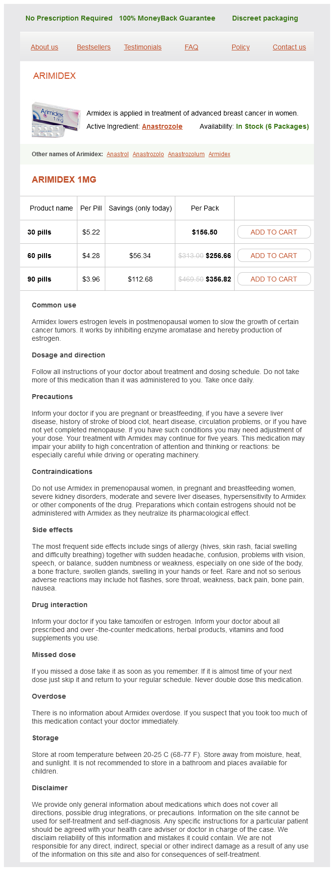
Arimidex dosages: 1 mg
Arimidex packs: 30 pills, 60 pills, 90 pills

At the hilum of the spleen women's health clinic king st london ontario arimidex 1mg discount, this layer passes on to the surfaces of the spleen lining (in that order) its gastric impression, the diaphragmatic surface and the renal impression, and thus returns to the hilum. In this way, the spleen comes to be lined all round by peritoneum except at the hilum. The spleen is separated from the diaphragm, from the kidney and from the stomach, by a part of the greater sac of peritoneum. However, the tail of the pancreas passes in the interval between the two layers of the lienorenal ligament and comes into direct contact with splenic tissue. The terminal part of the artery divides into a number of branches that pass through the lienorenal ligament to enter the hilum of the spleen. The splenic artery also gives off the short gastric and left gastroepiploic branches. The spleen receives autonomic nerves that reach it by running along the plexus surrounding the splenic artery. Accessory spleens (splenuneuli) may be present in structures near the organ including the gastrosplenic and lienorenal ligaments, the hilum of the spleen itself, the pancreas, and along the splenic artery. When enlarged considerably (to almost twice its normal size) the spleen projects from under the costal margin and can be felt on palpation of the abdomen. Because of the close relationship of the tail of the pancreas to the hilum of the spleen the former can be injured during splenectomy. Radio-opaque dyes can be introduced into the portal venous system through a needle introduced into the spleen (splenovenography or splenoportography). The technique has now been largely replaced by coeliac angiography during the venous filling phase. The arteries that supply the stomach, the intestines, the liver, the pancreas and the spleen are the ventral branches of the abdominal aorta. The veins draining these organs do not drain directly into the systemic circulation. After passing through the sinusoids of the liver the blood reaches the inferior vena cava through hepatic veins. The coeliac trunk arises from the front of the uppermost part of the abdominal aorta just below the aortic a opening in the diaphragm. On either side, the coeliac trunk is related to the corresponding crus of the diaphragm and to the coeliac ganglion. Chapter 29 Blood Vessels of Stomach, Intestines, Liver, Pancreas and Spleen 585 29. Here it lies in front of the neck of the pancreas and to the left of the bile duct. The gastroduodenal artery also gives off small branches to the stomach, the pancreas, and the duodenum.
However menstruation after tubal ligation discount 1mg arimidex with amex, it can pass forwards over the scrotum, over the penis, and upwards over the pubic symphysis into the lower part of the anterior abdominal wall. It cannot spread laterally into the thigh because of the attachment of the membranous layer to the ischiopubic rami. However, it can pass laterally over the lower part of the abdominal wall and can then pass downwards across the inguinal ligament into the thigh (25. The relationship of the abdominal viscera to the surface of the body is of considerable importance. As the anterior and lateral walls of the abdomen are devoid of skeletal landmarks (except at their upper and lower ends), reference has to be made to some imaginary planes. The abdomen can be divided into nine regions by using two transverse and two vertical planes which are as follows (25. This lies midway between the upper border of the manubrium sterni (suprasternal notch) and the upper border of the symphysis pubis. The vertical planes used for subdividing the abdomen into regions are the right and left lateral planes. On the anterior aspect of the body, they are represented by the right and left lateral lines. The upper end of each line is at the midpoint between the medial and lateral ends of the clavicle. Its lower end is midway between the anterior superior iliac spine and the pubic symphysis. The right and left lateral lines are commonly referred to as the midclavicular lines. Roughly, it can be said to lie at the level of the lower end of the body of the sternum. The lower limits of the abdominal cavity (excluding the true pelvis) are marked by the right and left inguinal ligaments. Keeping in mind the planes and limits defined above, the abdomen can be divided into the following nine regions (25. The subcostal plane is at the level of the lowest part of the costal margin (formed by the tenth costal cartilage). Some authorities use this plane (instead of the transpyloric) for dividing the abdomen into the regions mentioned above. When drawn on the posterior surface of the body this plane cuts the spine of vertebra l4, and is used as a guide to locate this spine. Its junction with the costal margin (9th costal cartilage) lies at the level of the transpyloric plane. The umbilicus is a prominent feature on the anterior abdominal wall, but is not a useful landmark because of variability in its position. In the healthy young adult it usually lies at the level of the intervertebral disc between l3 and l4.

The anterior aspect of the lower end of the humerus shows two depressions: one just above the capitulum and another above the trochlea menstrual vs estrous cycles buy arimidex 1mg low cost. Parts of the head of the radius and of the coronoid process of the ulna lie in these depressions when the elbowisfullyflexed. Another depression is seen above the trochlea on the posterior aspect of the lower end (2. This depression is called the olecranon fossa as it lodges the olecranon process of the ulna when the elbow is fully extended. The pectoralis major is inserted into the lateral lip of the intertubercular sulcus. Of the three insertions into the intertubercular sulcus that of the pectoralis major is the most extensive, and that of the latissimus dorsi is the shortest. The coracobrachialis is inserted into the rough area on the middle of the medial border. Thebrachialis arises from the lower halves of the anteromedial and anterolateral surfaces of the shaft. The pronator teres (humeral head) arises from the anteromedial surface, near the lower end of the medial supracondylar ridge. The brachioradialis arises from the upper two-thirds of the lateral supracondylar ridge. The extensor carpi radialis longus arises from the lower one-third of the lateral supracondylar ridge. The superficial flexor muscles of the forearm arise from the anterior aspect of the medial epicondyle. The lateral head of the triceps arises from the oblique ridge on the upper part of the posterior surface, just above the radial groove. The medial head of the muscle arises from the posterior surface below the radial groove. The upper end of the area of origin extends onto the anterior aspect of the shaft. On the medial side, the line of attachment dips down by about a centimetre to include a small area of the shaft within the joint cavity. The line of attachment of the capsule is interrupted at the intertubercular sulcus to provide an aperture through which the tendon of the long head of the biceps leaves the joint cavity. The capsular ligament of the elbow joint is attached to the lower end of the bone. Anteriorly the line of attachment reaches the upper limits of the radial fossa and the coronoid fossa. The medial and lateral epicondyles give attachment to the ulnar and radial collateral ligaments respectively. The intertubercular sulcus lodges the tendon of the long head of the biceps brachii.

The hip bone has three primary centres womens health associates buy arimidex 1 mg without prescription, one each for the ilium, the ischium and the pubis. At birth, the ilium, ischium and pubis are separated by a Y-shaped cartilage present in the region of the acetabulum. The inferior ramus of the pubis and the ramus of the ischium are at first separated by cartilage. These centres appear at about the age of puberty or later and fuse with the rest of the bone between 20 and 25 years of age. We have seen that the bony pelvis is made up of the two hip bones, the sacrum and the coccyx (9. It may be subdivided into the greater (or false) pelvis and the lesser (or true) pelvis. The walls of the greater pelvis are formed by the broad upper parts of the two iliac bones (iliac fossae), and posteriorly by the base of the sacrum. Note that the greater pelvis has no bony anterior wall, and that it is merely the lower part of the abdomen. The communication between the greater and lesser pelvis is called the superior pelvic aperture or pelvic inlet. Behind by the sacral promontory, and the ridge separating the superior and anterior surfaces of the sacrum b. The arcuate line, the pecten pubis and the pubic crest are collectively referred to as the linea terminalis. On either side by the pelvic surfaces of the ilium and ischium below the arcuate line c. Laterally, in that order, by the ischial tuberosity, the lesser sciatic notch, the ischial spine and the greater sciatic notch. When the ligaments are intact the lateral margins are formed by the sacrotuberous ligaments (that stretch from the side of the sacrum and coccyx to the ischial tuberosity). The anteroposterior diameter is measured from the upper border of the symphysis pubis to the sacral promontory. The oblique diameter is measured from one iliopubic eminence to the opposite sacroiliac joint. The anteroposterior diameter is measured from the apex of the coccyx to the lower border of the symphysis pubis. The oblique diameter is measured from the midpoint of the sacrotuberous ligament of one side to the junction of the ischial and pubic rami on the other side. Clinical Note Sex Differences in the Pelvis It is significant in forensic medice Of all the bones of the human skeleton sexual differences are most marked in the pelvis, and these are useful in deciding whether a given pelvis belongs to a male or a female individual. As a rule, the male pelvis is more strongly built than in the female, and the bones have more prominent muscular markings. All the articular areas including the acetabulum are larger in the male, for transmission of greater body weight. Some points that are really useful in deciding the sex of a given pelvis are as follows.
If there is an ulcer or cancer on the tongue (lingual nerve) menstruation length arimidex 1mg generic, the pain may again be felt over the ear and temple (auriculotemporal). In frontal sinusitis (sinus supplied by a branch from the supraorbital nerve) the pain is referred to the forehead (skin supplied by supraorbital nerve). In fact headache is a common symptom when any structure supplied by the trigeminal nerve is involved. A source of irritation in the distribution of the nerve may cause severe persistent pain (trigeminal neuralgia). In such cases pain can be relieved by injection of alcohol into the trigeminal ganglion, into one of the divisions of the nerve, or into its sensory root. In this connection it is important to know that the fibres for the maxillary and mandibular divisions can be cut without destroying those for the ophthalmic division. This is possible as the fibres for the ophthalmic division lie separately in the upper medial part of the sensory root. Finally, it may be noted that trigeminal pain can also be relieved by cutting the spinal tract of the trigeminal nerve: this procedure is useful especially for relieving pain in the distribution of the ophthalmic division as pain can be abolished without loss of the sense of touch and, therefore, without the abolition of the corneal reflex. The tip of the needle is now very near the inferior alveolar nerve, just before it enters the mandibular canal. The lingual nerve lies very close to the medial side of the third molar tooth, just deep to the mucosa. In cases of cancer of the tongue, having intractable pain, the lingual nerve can be cut at this site to relieve pain. Structures through which the nerve passes are shown as if transparent 908 Part 5 Head and Neck 1. It is attached to the brainstem by two roots: A large motor root, and a smaller sensory root. These roots are attached in the lateral part of the groove between the lower border of the pons and the upper border of the medulla (43. The sensory root is attached midway between the motor root (medially) and the vestibulocochlear nerve (laterally). From this attachment the motor and sensory roots pass forwards and laterally and leave the posterior cranial fossa by entering the internal acoustic meatus (on the posterior aspect of the petrous temporal bone). The nerve has a complicated course through the substance of the petrous temporal bone. This part of the nerve bears the genicular ganglion (so called because it is situated on a sharp bend of the nerve). It immediately enters the substance of the parotid gland and runs forwards within it and ends behind the neck of the mandible by dividing into several branches.
Although breast cancer jerseys buy arimidex 1mg mastercard, less detail is seen than in an X-ray film the technique allows a dynamic study. Computed Tomography and Magnetic Resonance Imaging these are newer techniques that provide images of the chest with great detail. They often help to establish diagnosis in many cases in which routine X-ray examination is inconclusive. The interior of the trachea and bronchi can be visualised through an instrument called a bronchoscope. More recent bronchoscopes, based on fibre optics, are flexible and can be passed into smaller bronchi. Apart from examining the interior of the bronchial tree, bronchoscopy can also be used to obtain bronchial secretions and tissue for examination, or to remove foreign bodies that may enter the bronchi. At the bifurcation of the trachea we can see the openings into the principal bronchi. Bronchography is a procedure in which X-ray pictures of the bronchi can be obtained after instilling a radioopaque substance into them. The techniques mentioned above give information about the structural status of the respiratory system. Of equal importance are investigations of function details of which can be found in books on physiology and of medicine. It is more likely to enter the right bronchus than the left as the right bronchus is wider, shorter and more vertical. Inflammations of the lungs and bronchi can be caused by bacteria, by viruses, by allergy, or by irritant action of pollutants in the atmosphere. In some cases serious lung infections cause localised necrosis of tissue leading to formation of a lung abscess. Most lung abscesses are caused by inhalation of infective material from infected sinuses (sinusitis), tonsillitis, or dental infection. Amongst bacterial infections of the lungs special mention needs to be made of pulmonary tuberculosis. The disease is now treatable and preventable, but it is still prevalent especially amongst populations that live in unhygienic conditions and are poorly nourished. Sensitivity to substances of plant, animal or chemical origin in the atmosphere can also be responsible for diseases that have a predominantly allergic basis. One such condition is bronchial asthma in which spasm of bronchial muscle causes considerable difficulty in breathing. Some degree of bronchospasm may also be present in various other conditions seen in the lungs. In some cases chronic respiratory obstruction leads to considerable dilatation of alveoli. Obstruction to a bronchus from any cause can lead to collapse of the part of lung supplied by it.
This surface is separated from the cavity of the wrist joint by an articular disc breast cancer 2b survival rate generic arimidex 1 mg overnight delivery. This surface articulates with the ulnar notch of the radius to form the inferior radioulnar joint. The styloid process is a small downward projection that lies on the posteromedial aspect of the head. Between the styloid process and the head the posterior aspect is marked by a vertical groove. It is of importance to note that in the intact body the tip of the styloid process of the ulna lies at a higher level than the styloid process of the radius. The Shaft the shaft of the ulna has a sharp lateral or interosseous border, and less prominent anterior and posterior borders. The upper part of the interosseous border is continuous with the supinator crest mentioned above. The lower part of this border is indistinct and ends on the lateral side of the head. The posterior border begins at the apex of the triangular area on the posterior aspect of the olecranon process (2. The anterior surface of the ulna lies between the interosseous and anterior borders. Its lower part shows an oblique ridge that runs downwards and medially from the interosseous border. The upper of these lines runs obliquely downwards and medially across the upper part of the surface. It starts at the posterior end of the radial notch and terminates by joining the posterior border. The part below the oblique line is subdivided into medial and lateral parts by a vertical ridge. The brachialis is inserted into the anterior surface of the coronoid process including the tuberosity. The triceps is inserted into the posterior part of the superior surface of the olecranon process (2. The flexor digitorum profundus arises from the upper three-fourths of the anterior and medial surfaces. The supinator arises from the supinator crest and from the triangular area in front of it. The flexor pollicis longus (occasional ulnar head) arises from the lateral border of the coronoid process. The flexor digitorum superficialis (ulnar head) arises from the tubercle at the upper end of the medial margin of the coronoid process. The pronator teres (ulnar head) arises from the medial margin of the coronoid process. The pronator quadratus arises from the oblique ridge on the lower part of the anterior surface of the shaft.
The posterior intercostal and subcostal arteries have already been described (Chapter 18) womens health hudson ny purchase 1mg arimidex fast delivery. These arteries supply the bronchi, the connective tissue of the lung and related lymph nodes. It may arise from the upper left bronchial artery or from the third right posterior intercostal artery. Other Arteries in the Thorax the internal thoracic artery, and the superior intercostal artery arise in the neck and descend into the thoracic wall. Veins that Drain the Heart these are the coronary sinus and its tributaries; and some small veins. Large Veins Present in the Mediastinum these are the superior vena cava, the right and left brachiocephalic veins, the inferior vena cava, and the pulmonary veins. Most of the veins draining the heart wall end in a wide vein, about two centimeters long, called the coronary sinus (21. The great cardiac vein is seen mainly on the sternocostal aspect of the heart (21. It ascends in the anterior interventricular groove (parallel to the anterior interventricular branch of the left coronary artery). Veins not normally seen from the front are drawn as if the walls of the chambers of the heart were transparent 21. Veins not normally seen from the back are drawn as if the walls were transparent 2. It receives a large left marginal vein which corresponds to the left marginal branch of the left coronary artery. The small cardiac vein is situated at the junction of the base of the heart and its diaphragmatic surface (21. The small cardiac vein receives the right marginal vein which is seen on the sternocostal surface just above the inferior border of the heart. Its position thus corresponds to that of the right marginal branch of the right coronary artery. The middle cardiac vein begins near the apex of the heart and runs backwards on the diaphragmatic surface (21. The posterior vein of the left ventricle runs backwards on the diaphragmatic surface of the ventricle and ends in the coronary sinus. In addition to the above some anterior cardiac veins lying on the right ventricle open into the right atrium. A number of small venae cordis minimae drain directly into the chambers of the heart.
Bozep, 35 years: The region where the front of the arm becomes continuous with the front of the forearm is marked by a triangular depression called the cubital fossa.
Boss, 64 years: The lower end has to be marked at the level of the lower border of the twelfth vertebra.
Tufail, 47 years: Neutropenic or lymphopenic patients do not have impaired wound healing, whereas macrophage-deficient (quantity or function) patients heal poorly 1, A; 2, B; 3, C.
Emet, 34 years: In this position the fluid gravitates into the pelvis where absorption is much less pronounced (see rectouterine pouch, below).
References